Proteomics In Motion: Visualizing Cilia-Related Processes At The Molecular Level
In recent years, proteomics has become one of the most important tools for understanding how organisms work at a molecular level. Proteomics is used to identify and quantify proteins in biological samples, allowing us to understand the complex processes that occur inside cells. In this article, I’ll be exploring how proteomics can help us visualize cilia-related processes at the molecular level.
Cilia are small hair-like structures found on many types of cells throughout our bodies and play an essential role in numerous biological functions such as respiration, movement and sensation. Using proteomics we can now look more closely into these processes at a cellular level and gain further insight into their complexities.
Using state of the art technology, researchers have developed new methods for visualizing cilia-related events within individual cells. By combining powerful microscopes with sophisticated protein analysis techniques, scientists can observe changes in both structure and function over time – giving us unprecedented access to the inner workings of these tiny yet vital organelles!
Overview Of Proteomics
I’m going to take you on a journey through proteomics, the technology that is allowing us to gain unprecedented insight into cilia-related processes at the molecular level. Proteomics involves studying proteins and their interactions with one another for better understanding of biological systems or organisms. Through this method we can identify new pathways, biomarkers, and drug targets which can be used in further research studies.
The combination of cutting edge technologies such as Mass Spectrometry (MS) and Liquid Chromatography (LC), along with sophisticated software and data analysis techniques, allow us to visualize these cilia-related processes down to the molecular level. With this knowledge, we are able to investigate how various elements within cells interact with each other in order to carry out important functions related to cell development, growth and homeostasis.
To move forward in our quest for greater insights into these processes, it is important that we understand not only what molecules are present but also how they interact with one another. That’s where proteomics comes in – providing an avenue towards uncovering previously unseen secrets about life at its smallest scale. From here, let’s explore how cilia-related processes are visualized using proteomic technology.
How Cilia-Related Processes Are Visualized
Now that we’ve discussed proteomics, let’s move on to how cilia-related processes are visualized. Proteomics visualization is the act of using various techniques and methods to visualize cilia-related processes at a molecular level. This type of visualization can help researchers gain insight into how these small structures work together in order to carry out their functions.
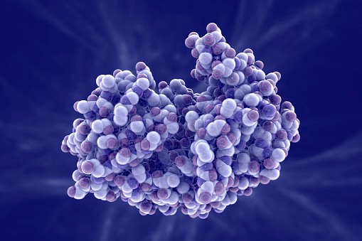
Visualizing cilia-related processes can be done through several different methods. For example, scientists use microscopy imaging technology such as fluorescence or confocal microscopy to observe proteins within cells. Additionally, they may utilize spectroscopic techniques like mass spectrometry to analyze protein composition and structure. Finally, X-ray crystallography or nuclear magnetic resonance (NMR) could be employed for detailed structural information about proteins and other molecules. All of these visualization techniques offer unique insights into the workings of cilia-related processes at the molecular level.
These visualization tools allow us to have an unprecedented view into how these tiny organelles interact with each other and function in our bodies. Through this knowledge, researchers can develop better treatments for diseases related to malfunctioning cilia, including cystic fibrosis and Kartagener syndrome. It also provides key information on understanding human development more broadly by shedding light on how crucial cellular components are connected and regulated during embryonic growth and development.
Benefits Of Molecular Visualization
The use of molecular visualization to analyze cilia-related processes has many benefits. Proteomics specialists can gain a detailed understanding of these processes at the molecular level, and thus be better equipped to make informed decisions about treatments for diseases associated with cilia-related abnormalities.
Molecular visualization techniques allow us to:
- Visualize protein structures in 3D
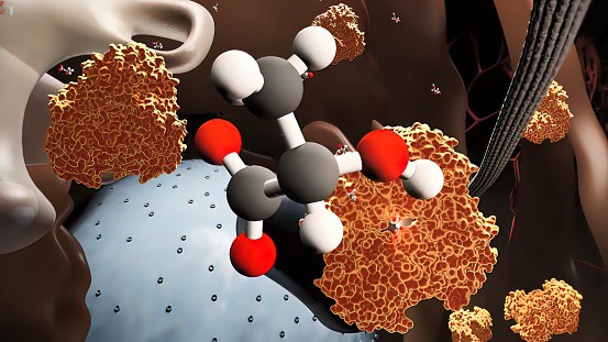
- Analyze how proteins interact with one another
- Understand how mutations affect gene expression
- Identify potential drug targets
- Investigate how drugs act on specific molecules or pathways
These techniques enable us to explore complex biological systems in ways that were not possible before, accelerating our ability to develop new treatments for various conditions related to cilia-related abnormalities. Molecular visualizations also help scientists more accurately identify important cellular components and understand their roles within the cell, giving us crucial insights into disease mechanisms. Overall, molecular visualization is a powerful tool that helps proteomics specialists uncover valuable information about cilia-related processes at the molecular level.
Conclusion
Proteomics has revolutionized our understanding of cilia-related processes. By being able to visualize them at the molecular level, we can gain a deeper insight into how these processes operate and interact with each other. Through this visualization process, researchers are now able to make groundbreaking discoveries that would have otherwise remained inaccessible.
Visualizing cilia-related processes at the molecular level is revolutionary because it allows us to look back in time and see what might have been missed or overlooked when trying to understand complex biological systems. The ability to “rewind” gives us an unprecedented opportunity to dive deep into the natural world and uncover insights previously unknown. I believe this technology will empower us like never before, as we continue pushing boundaries in proteomics research.
By leveraging proteomic technologies such as visualizing cilia-related processes at the molecular level, we can unlock science’s greatest mysteries and bring about solutions for some of humanity’s most pressing challenges. We all stand on the precipice of a new era – one where science fiction becomes reality!
